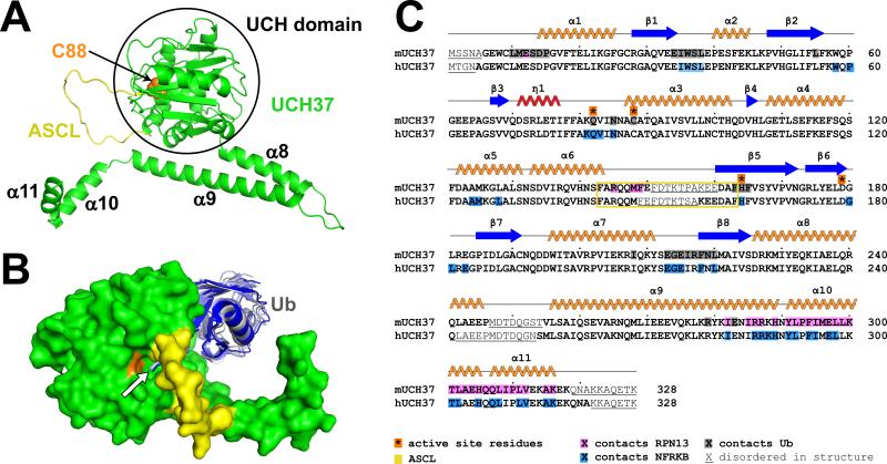
Figure 1. Structure of UCH37 and ubiquitin complex.
(A) UCH37, ribbon representation, as seen in the RPN13 complexes. The catalytic C88 is shown as orange spheres; active site crossover loop (ASCL), yellow. Helices α8-α11 that comprise the CTD are labeled.
(B) Surface representation of UCH37 with ubiquitin (blue-gray) from the RPN13 complex and structures of ubiquitin in complex with other UCH family members (Boudreaux et al., 2010; Johnston et al., 1999; Misaghi et al., 2005; Morrow et al., 2013) following superposition on the UCH domains. The ubiquitin C-terminus is indicated with an arrow.
(C) Sequence and secondary structures of murine UCH37 isoform 2 (RPN13 and ubiquitin complex) and human UCH37 isoform 2 (NFRKB complex).

