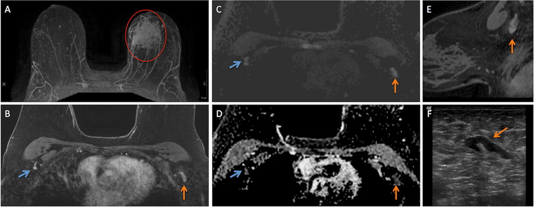
Figure 1.
57 year-old woman presented with a 26 mm invasive ductal carcinoma (IDC) in the left breast for a bilateral breast MRI to assess extent of disease with dynamic contrast enhanced (DCE) and diffusion weighted imaging (DWI) techniques.
A. Regional non-mass enhancement (red circle) is present in the left breast on maximum intensity projection (MIP) image, site of biopsy-proven IDC (red circle) in the left breast.
B. Initial phase post-contrast T1-weighted image with fat suppression demonstrates a morphologically suspicious level 1 axillary lymph node (ALN) in the left axilla (vertical orange arrow) and a representative contralateral morphologically normal level 1 ALN (angled blue arrow) in an analogous location.
C. Both the suspicious ALN (vertical orange arrow) and normal ALN (angled blue arrow) exhibit high signal intensity on DWI.
D. Both the suspicious ALN (vertical orange arrow) and normal ALN (angled blue arrow) are dark on ADC map (D) confirming the presence of restricted diffusion.
E. Sagittally-oriented contrast-enhaced T1-weighted, fat suppressed reformations demonstrates the morphology of the suspicious ALN (vertical orange arrow) in the breast ipsilateral to the IDC.
F. Morphology of the suspicious ALN on ultrasound (angled orange arrow) that ultimately underwent ultrasound-guided biopsy correlates well with morphological appearance on MRI (B, E).

