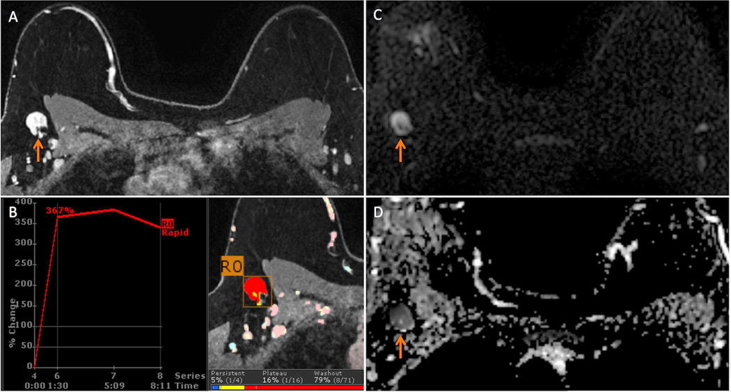
Figure 3.
40 year-old woman with newly diagnosed high nuclear grade ductal carcinoma in situ (DCIS) spanning 50 mm in the right breast presents for bilateral breast MRI to evaluate extent of disease. A suspicious and ultimately biopsy-proven benign (on both core needle biopsy and sentinel lymph node biopsy) ipsilateral axillary lymph node (ALN) was identified on the MRI. The asymmetrical size and suspicious morphology of this lymph node was presumed to be due to reactive changes from recent ipsilateral breast biopsy performed 1 week prior to the MRI.
A. T1-weighted fat-suppressed contrast enhanced axial image demonstrates the suspicious ALN (arrow) measuring 29 mm in maximal size and exhibiting diffuse cortical thickening and loss of fatty hilum.
B. The suspicious ALN exhibited 308% initial peak enhancement with 83% delayed washout (red color overlay) on computer aided evaluation.
C. On diffusion weighted imaging (DWI), the ALN exhibits high signal on the b=600 s/mm2 image (orange arrow).
D. The suspicious ALN is dark on the apparent diffusion coefficient (ADC) map (arrow, mean ADC = 0.78 × 10⁻³ mm2/s), indicating restricted diffusion.

