
Figure 1.
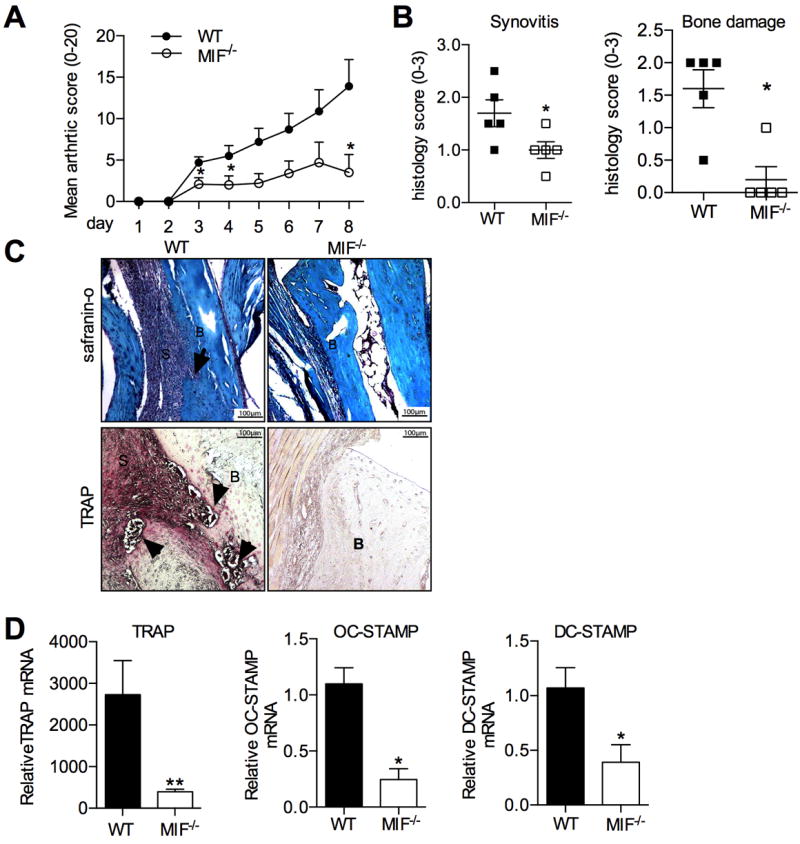
Inflammatory arthritis in WT and MIF-/- mice with K/BxN serum transfer arthritis. A: Clinical scoring of joint inflammation in K/BxN serum-injected WT mice (closed circles) and MIF-/- (open circles). B: Histological scores for synovitis (left) and bone damage (right) in WT and MIF-/- mice. C: Top: Safranin O stained sections of ankles WT and MIF-/- mice with K/BxN serum transfer arthritis, showing synovitis (S) and bone erosion (arrows) (B:bone). Bottom: TRAP histochemical staining (pink) in ankle sections of WT and MIF-/- mouse (‘B’=bone, ‘S’=synovium and TRAP+ cells indicated by arrows). Original magnification x200. C: Osteoclast-associated gene expression determined using RNA extracted from ankles of WT and MIF-/- mice with K/BxN serum transfer arthritis. All values were expressed as mean ±SEM; each group contains 5 mice. *p<0.05, **p<0.01, MIF-/- compared to WT.