
Figure 5.
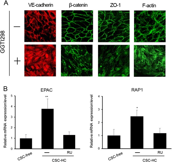
Impairment of hydrocortisone-mediated facilitation of adherens junction formation by inhibiting RAP1 activity. (A) Immunocytochemistry was performed to examine the cellular localization of VE-cadherin, β-catenin, and ZO-1 in cells cultured with CSC-HC in the presence or absence of a RAP1 inhibitor, GGTI298 (5 μM). Phalloidin staining was performed to analyze F-actin distribution. The experiments were repeated three times and representative results are shown. The “+” or “-” symbols indicate the addition of GGTI298 or DMSO (0.1%) to the medium, respectively. (B) EPAC and RAP1 mRNA expression levels in cells cultured with CSC-free or CSC-HC were examined by qPCR. Effect of RU486 on HC-mediated induction of mRNA expression was also investigated. mRNA expression levels were calculated relative to the value obtained from CSC-free cells (set as the basal level =1). Each value is expressed as mean ± S.D. obtained from three independent experiments. The single and double asterisks indicate represent p < 0.05 and p < 0.01 respectively, relative to CSC-free.