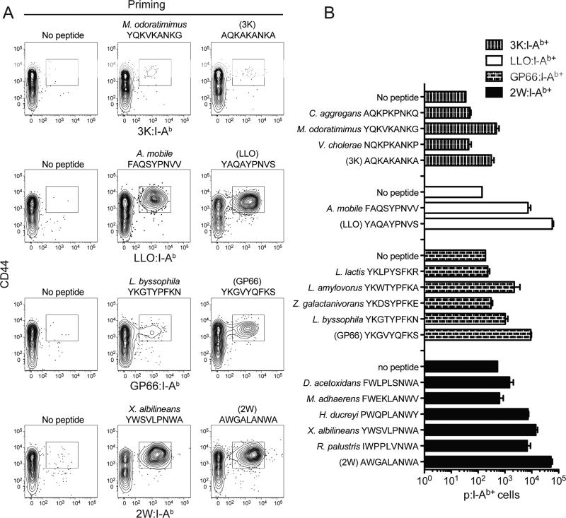
Figure 3. Conservation of TCR contact amino acids predicts p:I-Ab cross-reactivity.
(A) Contour plots showing parent p:I-Ab tetramer staining (indicated on the Y-axes) of spleen and lymph node cells from mice primed 11 days earlier with CFA alone (no peptide), homologous bacterial peptide, or parental peptide. CD44hi tetramer-binding cells are indicated in the rectangular gates.
(B) Average number of 3K:I-Ab+, LLO:I-Ab+, GP66:I-Ab+, or 2W:I-Ab+ CD4+ T cells 11 days after priming with the indicated peptides emulsified in CFA (± SEM, n = 3 mice) from 1 experiment, which was representative of another.

