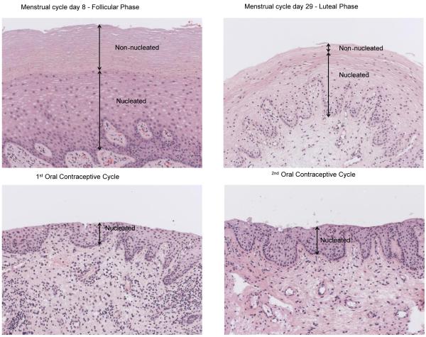
Figure 3. Vaginal epithelial changes following COC administration.
Hematoxylin and eosin stained vaginal biopsies from BB967 during follicular (day 8), luteal (day 29), and first and second medicated phases. The follicular phase biopsy shows an overall thicker vaginal epithelial thickness, with a markedly thickened non-nucleated layer compared to the luteal phase biopsy. Both biosies taken during COC administration show a nucleated layer that is thinner than both follicular and luteal phase specimens, and complete absence of a non-nucleated layer.

