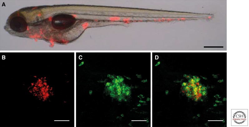
Figure 4.
Pathology in embryos. (A) Merged bright-field and fluorescent image of a zebrafish embryo infected with red fluorescent M. marinum and photographed at 5 dpi. (Adapted with permission from Stoop et al. 2011.) Clustering of mycobacteria and early granuloma formation is shown as red spots. (B–D) Higher magnification of an early granuloma at 5 dpi formed after bloodstream infection, derived from analysis using confocal imaging by our research group. (B) M. marinum E11 (in red), (C) phagocytes stained with anti-L-Plastin (in green), (D) merge of B and C confirming the colocalization of these cells in early granulomas in zebrafish embryos. Scale bar, 35 µm.

