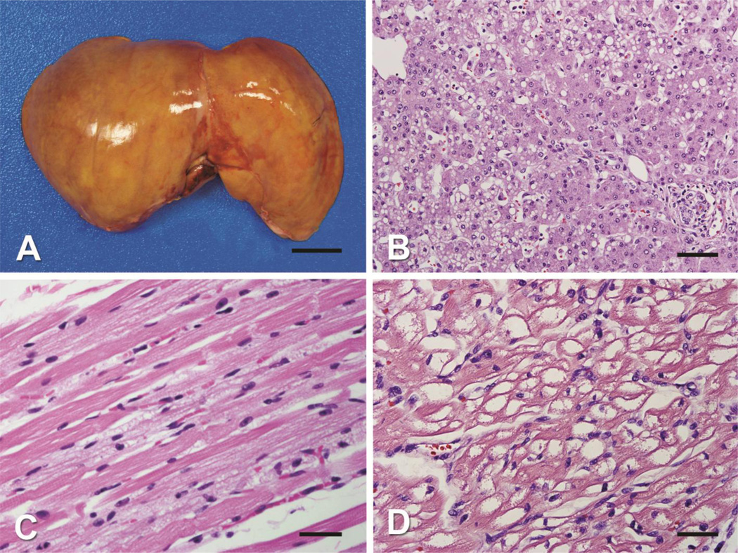
Figure 1. Pathology of tissues of HSD10 patient.
A) A gross photograph of the enlarged liver shows diffuse yellow discoloration (scale bar = 2.5 cm). B) The hepatocytes in zone 2 show micro- and macrovesicular steatosis (H&E stain, original magnification 40X objective, scale bar 100 µm). C) The skeletal muscle shows numerous cytoplasmic vacuoles (H&E stain, original magnification 40X objective, scale bar = 35 µm). D) The cardiomyocytes show extensive cytoplasmic vacuoles (H&E stain, original magnification 40X objective, scale bar = 35 µm).

