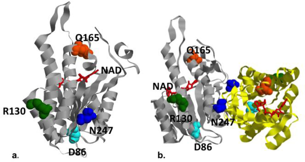
Figure 6. Modeling of human HSD10/MRPP2 mutations.
Locations of mutated residues in the HSD10/MRPP2 protein, and protein dimmer Crystal structure of human HSD10 (pdb: 2023) with the locations of mutated residues highlighted. a.) The HSD10 patient’s mutation, Asn247, (shown in blue) is located on the same side of the enzyme as Arg130 (green) and Asp86 (light blue), away from the NAD (red) binding cleft and Gln165 (orange). b.) The mutations associated with a more severe phenotype (N247 and D86) are adjacent to the homodimerization domain (the third chain of the homodimer is shown in yellow ribbon).

