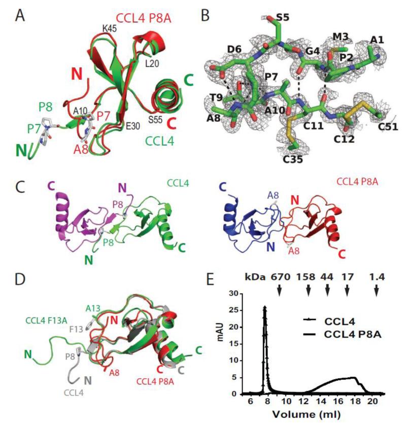
Figure 4. Structure of CCL4 P8A.
(A) Secondary structure comparison of CCL4 P8A (red) with a CCL4 monomer in the CCL4 polymer structure (green, PDB: 2X6L). Residues 7 and 8 are depicted as sticks. (B) A detailed hydrogen bond network at the N-terminal of CCL4 P8A to prevent its extension and subsequent dimerization. A composite omit 2mFo-DFc map was calculated in PHENIX and contoured at 1σ. (C) Comparison of the CCL4 dimer (top) with the hypothetical model of the CCL4 P8A dimer (bottom) to reveal the steric clash at the N-terminal end of CCL4 P8A in the model of CCL4 P8A dimer. (D) Size exclusion chromatography profile of CCL4 and CCL4 P8A. Chemokines (100 μl 1 mg/ml) were analyzed on Superdex 200 column with buffer containing 20 mM NaCl, 50 mM Tris (pH 7.8) at 4°C. The arrows indicate peak positions of the molecular weight standard. (E) Secondary structure comparison of CCL4 P8A (red, 3TN2), CCL4 F13A (green, 1JE4) and CCL4 (gray; 1HUM). Residues 8 and 13 were depicted as stick. Oxygen, nitrogen, sulfur and carbon atoms are shown in red, blue, yellow and gray, respectively.

