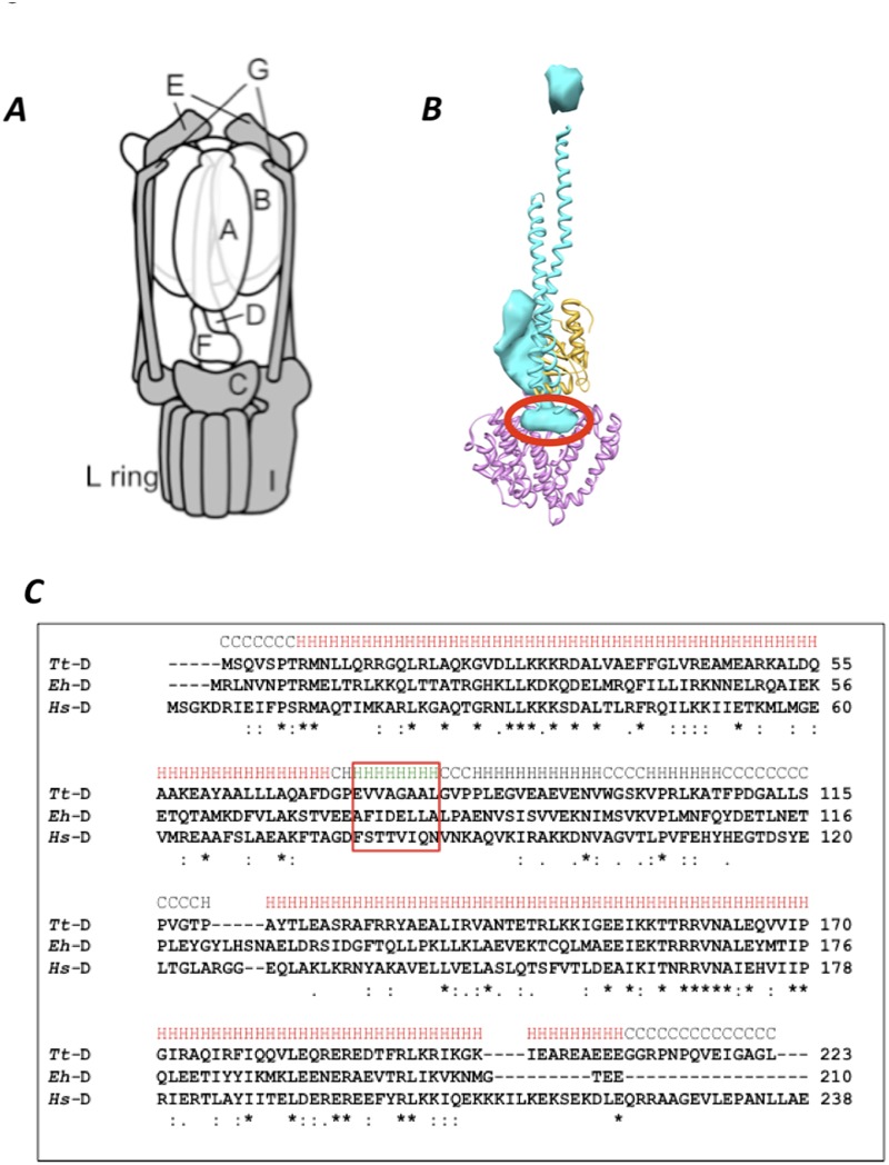
Fig 1. VoV1 and the short helix of V1-D subunit.
A, Schematic representation of T. thermophirus VoV1 [16]. Subunits in Vo and V1 are shown in gray and white, respectively. B, The structure of the central rotor apparatus of VoV1 obtained by EM density map (PDBID; 3J0J) with the short helix in V1-D subunit circled in red. The V1-D, V1-F and Vo-C subunits are represented in blue, yellow, and pink, respectively. C, Sequence alignment of V1-D subunit of T. thermophilus (Tt), E. hirae (Eh) and H. sapiens (Hs). Identical amino acid residues are represented by asterisks. The sequences of the short helix of the V1-D subunit are surrounded by the red box.

