
Figure 1.
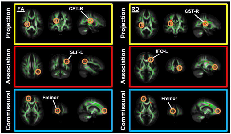
Results from TBSS analyses showing differences in fractional anisotropy (FA) and radial diffusivity (RD) in bipolar patients (BD) versus healthy controls (HC). Voxels are superimposed on the white matter skeleton (green). The background images are MNI152 template (MNI - Montreal Neurological Institute). Yellow-red clusters represent abnormalities in FA and RD in commissural, association and projection fibers (FDR-corrected p<0.01). Abbreviations: CST-R: right corticospinal tract, SLF-L: left superior longitudinal fasciculus, IFO-L: left inferior fronto-occipital fasciculus, Fminor: forceps minor.