
Figure 1.
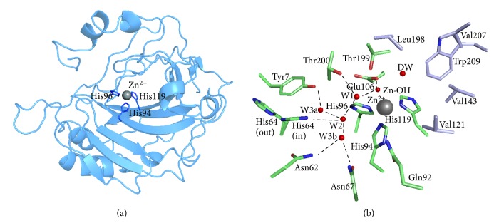
Structure of CA II. PDB ID: 3KS3. (a) Ribbon diagram depicting the overall structural fold. The active site zinc ion and coordinated histidines shown. (b) Active site and ordered waters (red spheres). Also shown are the hydrophilic (green) residues as well as the hydrophobic (purple) residues lining the active site.