
Figure 2.
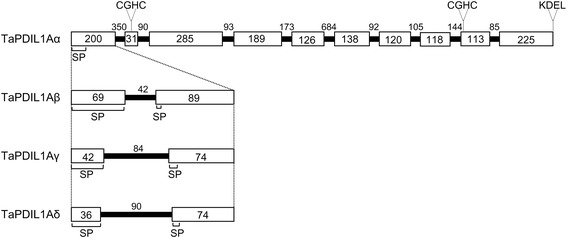
Splicing sites in TaPDIL1Aα , TaPDIL1Aβ , TaPDIL1Aγ , and TaPDIL1Aδ . Open boxes indicate exons, and solid black lines denote introns. The numbers indicate the size of each exon and intron (bp). The positions of the signal peptide (SP), the two CGHC motifs, and the C-terminal KDEL sequence are indicated.