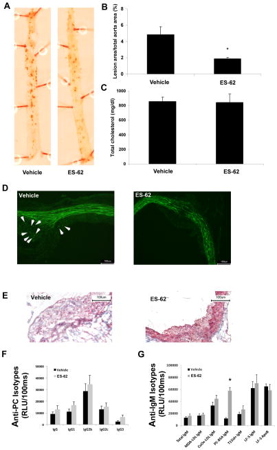
Fig. 2.
Acanthocheilonema viteae ES-62 treatment decreases atherosclerosis in gld.apoE−/− mice and not through induction of protective cross-reactive anti-phosphorylcholine (PC) antibodies. (A) Representative photographs and (B) quantification of Oil Red O-stained aortae from mice maintained on high cholesterol Harlan-Teklad Research Western diet for 12 weeks and treated with ES-62 or vehicle (*, P <0.01). (C) Quantification of total serum cholesterol, determined by a microtiter procedure according to the manufacturer’s instructions (Wako Diagnostics, Japan). Representative images of immunohistochemical analysis of atherosclerotic lesion by (D) F4/80 staining for macrophages (Caltag Laboratories, United Kingdom) and (E) trichrome staining for collagen shown within the lesion in blue. (F,G) Microtiter wells were coated with either antigens (malondialdehyde oxidized low density lipoprotein (MDA-LDL), copper oxidized LDL (CuOx-LDL), PC-BSA) or antibodies (AB1-2) at 2–5 μg/mL and serum antibodies to respective antigens were determined at different dilutions as described previously (Chang et al., 1999) (F) Serum levels of IgG isotypes binding to PC-BSA were determined by chemiluminescent ELISA. (G) IgM levels to indicated antigens as well as T15id+ IgM immune complexes were measured. Plasma levels of T15id+ natural IgM antibodies (EO6) were determined using the anti-T15-idiotypic monoclonal antibody AB1-2 (a mouse IgG1, which is absolutely specific for both the canonical T15 VH and the T15 VL regions) for capture, followed by detection steps using an anti-mouse IgM antibody. LF5 is a monoclonal antibody against mouse Apolipoprotein B (ApoB) and was used to capture ApoB containing particles on an ELISA plate. It was a kind gift from Stephen Young (University of California-Los Angeles, USA). IgM associated with LF5-ApoB particles was measured using anti-mouse IgM to detect IgM-ApoB immune complexes. In parallel wells, the amount of captured ApoB particles was measured using a commercial goat-anti ApoB to determine whether equal amounts of ApoB were captured in the assay. Values are given as relative light units (RLU) per 100 ms and represent the mean of triplicate determinations. Data are shown as mean ± S.E.M. values of all mice in each group (vehicle, n = 8; ES-62-treated, n = 9). The significant differences were assessed by a Mann Whitney test. P < 0.05 was considered significant.

