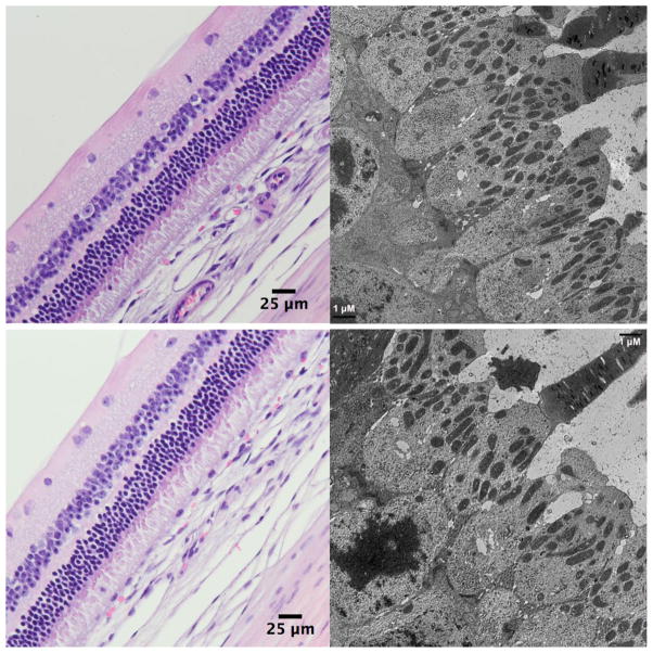
Figure 4.
Light microscopic images (left) and electron microscopic images (right) of the injected eye (upper panels) and its contralateral non-injected eyes (lower panels), showing comparable normal retinal layers under light microscope (62.5X) and normal ultrastructures of the photoreceptors under transmission electron microscope (3000X) at 4 weeks after the injection.

