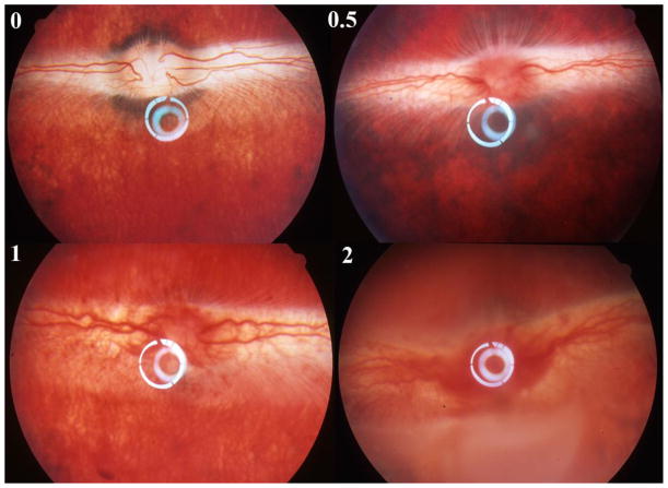
Figure 5.
Natural course of HSV-1 rabbit retinitis. Upper-left panel, normal rabbit fundus; Upper-right panel, retinitis grade 0.5 that is characterized by the hyperemia of optic nerve and no obvious signs of hemorrhage yet; Bottom-left panel, retinitis grade 1 that is characterized by the hemorrhages of the optic nerve and the medullary ray but no obvious retina infection (whitening foci) yet. The vitreous is still clear at this stage. Bottom-right, retinitis grade 2 is characterized by increased vitritis with decreased vitreous clarity as well as presence of discrete white patches of necrosis involving the inferior retina.

