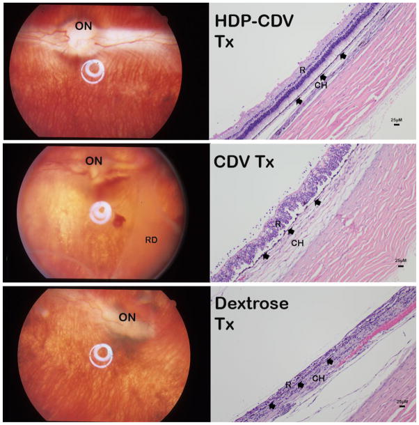
Figure 7.
Top-panels: HDP-CDV treated eye (HDP-CDV Tx) presented relatively normal fundus (left image); histology revealed only mild vitritis with inflammatory cells seen along the retina surface (right image). Mid-panels: CDV treated eye (CDV Tx) showed hazy vitreous with extensive retinal detachment (RD) (left image); histology revealed disorganized retina (R) layers with much more inflammatory cells along the retina surface (right image). Both RPE (arrowheads) and choroid (CH) are swelling. Bottom-panels: Dextrose injected eye revealed distorted optic nerve (ON) and shortened right side medullary ray (left image); histology showed no recognizable retinal layers and RPE was barely recognizable (arrowheads) with inflammatory cells packed in both retina area (R) and in Choroid (CH).

