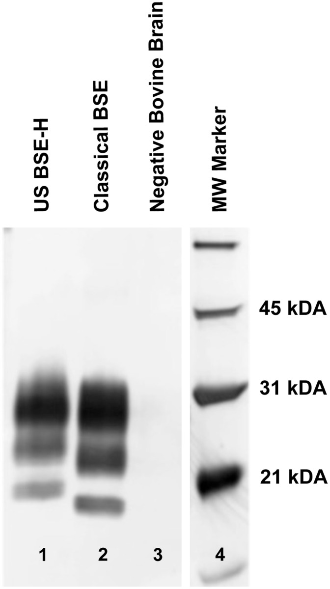
Fig 1. Western blot migration patterns of BSE-H and classical BSE.
Immunoblotting for PrPSc reveals the three characteristic glycoforms. A proteinase K-digested brain homogenate sample from an animal inoculated with H-Type BSE (lane 1) compared to a brain sample from an animal inoculated with classical BSE (lane 2) illustrates the higher molecular weight most noticeable in the BSE- H unglycosylated band. In a brain sample from a negative control animal (lane 3), the proteinase K pre-treatment destroys all antigenicity. Lane 4 contains molecular weight markers.

