
Figure 1.
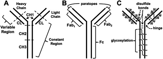
Simplified two-dimensional schematics of immunoglobulin G (IgG). A. Schematic showing the variable and constant regions of the IgG molecule for the heavy (CHx) and light chains (CL); B. Schematic of the antigen binding fragments (Fab1,2) and the fragment crystallizable region (Fc) of the IgG molecule; C. Schematic portraying intra- and interchain disulfide bonds as well as glycosylation of the Fab, Fc regions, and paratope antigen binding regions of the IgG molecule.