
Figure 4.
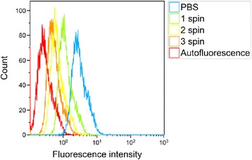
Flow cytometry histograms illustrating the effect of washing on cellular Nile Red fluorescence after incubation with nanoparticles. After incubation of 3 h at 37°C, the cells were rinsed 3 times with PBS and centrifuged and resuspended in medium 0 to 3 times.