
Figure 7.
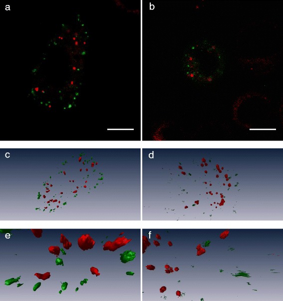
CLSM images of early endosomes labelled in cells incubated for 30 min with nanoparticles with encapsulated Nile Red (a) or with free Nile Red in medium (b). Below are 3-dimensional images generated from the CLSM z-stacks (c and d) together with zoomed in images of some vesicles (e and f). Early endosomes are labelled with CellLight Early Endosomes-GFP, which is shown in green, Nile Red is shown in red. A 488 nm laser was used to excite CellLight GFP, and GFP fluorescence was detected at 500–530 nm, while Nile Red was excited at 540 nm with detection at 550–640 nm. Scale bars are 10 μm.