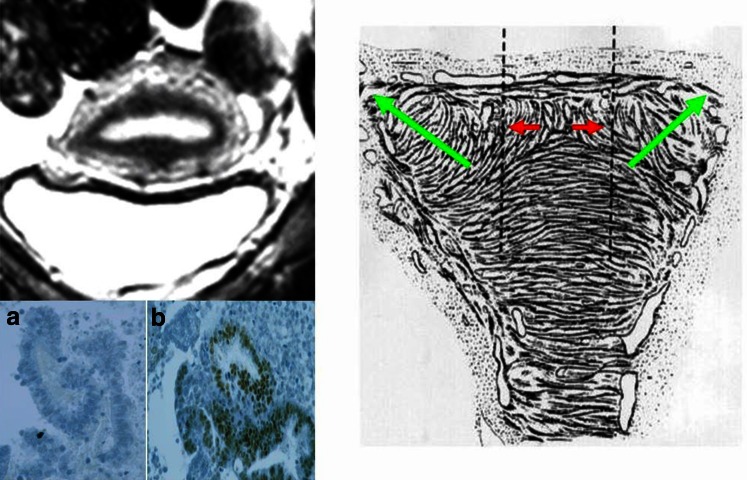
Fig. 10.
Schematic representation of the mechanism of uterine auto-traumatization by uterine peristalsis and hyperperistalsis at the fundo-cornual raphe. Green arrows direction of sperm transport. Red arrows distraction of basal stromal cells and archimyometrial myocytes at the fundo-cornual raphe by uterine peristalsis. With the development of an early adenomyotic lesion in the midline of the upper uterine corpus, a chronic process of proliferation and inflammation is established that facilitates the detachment of the basal endometrium. Fragment of detached functionalis (a) and a fragment of detached basalis (b) in menstrual blood (modified from [2, 4, 7])

