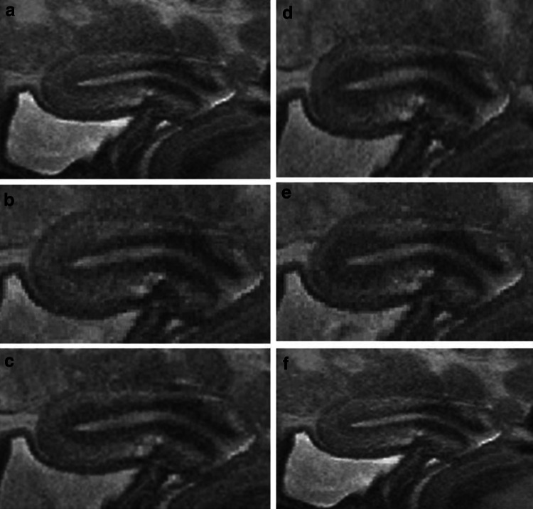
Fig. 5.
The course of a peristaltic wave of the archimyometrium as shown by a sequence of MRI scans obtained from cinematographic MRI scan in a healthy woman in the late follicular phase. Initially, the archimyometrium appears to be relaxed, indicated by a thin JZ with a less marked hypointensity (a). The peristaltic wave starts with tension of the archimyometrium in the lower half of the uterine corpus, indicated by marked hypointensity of the JZ (b). The zone of increased tension (marked hypointensity) moves in a fundal direction. A muscular package is built up, indicated by the rapid increase of the JZ as the wave moves in a fundal direction (c–e) followed by a rapid relaxation (f)

