Figure 6.

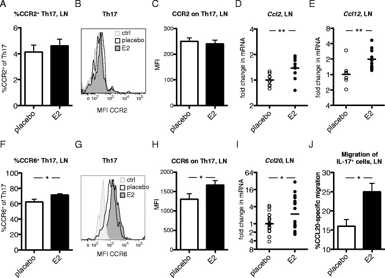
Th17 chemokine receptor expression and corresponding lymph node chemokine expression after estrogen treatment in collagen-induced arthritis. DBA/1 mice were ovariectomized, subjected to collagen-induced arthritis (CIA) and treated with 17β-estradiol (E2; 0.83 μg/day) or placebo. (A–C) Frequency of C-C chemokine receptor 2-positive (CCR2+) cells of T helper 17 (Th17) cells (A) and median fluorescence intensity (MFI) of CCR2 on Th17 cells (B,C) in lymph nodes (LNs) from CIA mice (day 14, single experiment with n = 10 or 11 mice/group). (B) Representative fluorescence-activated cell sorting (FACS) analysis plot where fluorescence minus one (FMO) is control. (D,E) mRNA expression of Ccl2 (D) and Ccl12 (E) in LNs of CIA mice (day 14, single experiment, n = 9 or 10 mice/group), determined by real-time quantitative PCR. (F–H) Frequency of CCR6+ cells of Th17 (F) and MFI of CCR6 on Th17 cells (G,H) in LNs of CIA mice (day 14, single experiments, n = 9 or 10 mice/group). (G) Representative FACS plot where FMO is control. (I) mRNA expression of Ccl20 in LNs of CIA mice (day 14, data pooled from two experiments, n = 20 mice/group). (J) CCL20-specific migration of interleukin (IL)-17+ cells. LN cells from mice with CIA (day 35) were put on a transwell assay, with CCL20 in the lower chamber, and the frequency of migrated cells was evaluated with an IL-17 ELISPOT assay. Data are representative of two independent experiments (n = 3 mice/group). To improve normal distribution of data, some data were log-transformed prior to statistical analysis (A, C, F and H). Bars or lines show arithmetic mean ± SEM (A, C, F, H and J) or geometric mean (D, E and I). Student’s t-test (A, C–F, H and J) or analysis of covariance with experiment as covariate (I) was used (*P <0.05, **P <0.01).
