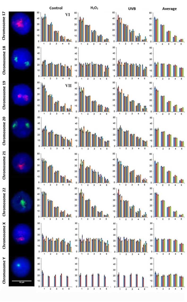
Fig 4. Radial distribution for chromosomes 17–22, X and Y in six subjects in unexposed, H2O2 and UVB exposed lymphocytes.
Fig. 4 displays representative FISH images and the radial distribution for CTs 17–22, X and Y. As in Figs. 2 and 3 moving from left to right the chromosome number is indicated followed by a representative FISH image for the CT and four histograms. Each histogram displays the proportion of fluorescence (%) from the nuclear interior toward the nuclear periphery (left to right). The first, second and third histogram displays the radial distribution for each CT in control, H2O2 and UVB exposed lymphocytes, respectively for each of the six subjects (1 to 6, left to right), with the exception of CTY which only contains data from the two male subjects enrolled in this study (subjects 1 and 2). The fourth histogram displays the average radial distribution for the enrolled subjects in unexposed (blue), H2O2 (red) and UVB (green) exposed lymphocytes. Error bars represent the standard error of the mean (SEM). Roman numerals indicate significant inter-individual variations in radial distributions (p<0.05) between subjects in control lymphocytes: VI- CT17 subject 6 (different to subject 5); and VII- CT19 subject 6 (different to subjects 1, 2, 3, 4, and 5).

