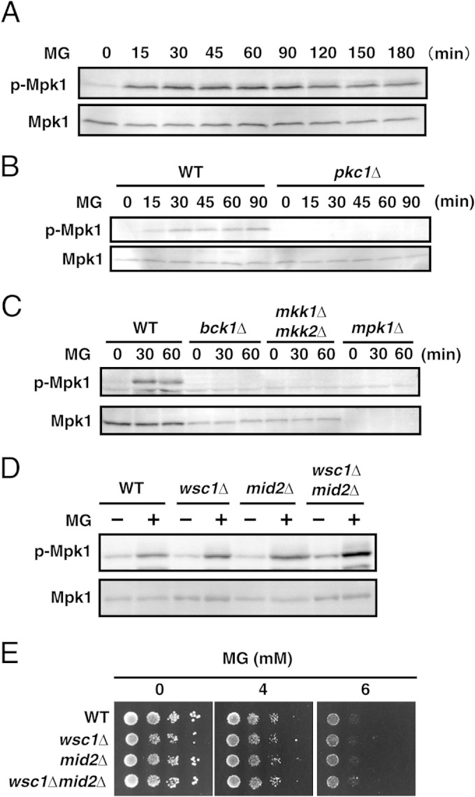
FIG 2.

Effects of MG on activation of the Mpk1 MAP kinase cascade. (A) Cells (YPH250) were cultured in SD medium until the A610 was 0.3, and 10 mM MG was added. The levels of phosphorylation of Mpk1 (p-Mpk1) and the Mpk1 protein (Mpk1) were determined after incubation for the prescribed times. (B) Wild-type (DL100) and pkc1Δ (DL376) cells were cultured in SD medium containing 1 M sorbitol until the A610 was 0.3 to 0.5 and were treated with 10 mM MG for the prescribed times. (C) Cells (YPH250) defective in the components of the Mpk1 MAP kinase cascade (bck1Δ, mkk1Δ mkk2Δ, and mpk1Δ cells) were cultured in SD medium containing 1 M sorbitol until the A610 was 0.3 to 0.5, and 10 mM MG was added. (D) Wild-type (YPH250), wsc1Δ, mid2Δ, and wsc1Δ mid2Δ cells were cultured in SD medium until the A610 was 0.3 to 0.5 and treated with 10 mM MG for 30 min, and the phosphorylation of Mpk1 was then determined. (E) The cells of each mutant in the YPH250 background were cultured in SD medium until the log phase of growth and serially diluted (1:10) with a 0.85% NaCl solution, and 4 μl of each cell suspension was then spotted onto SD agar plates containing MG.
