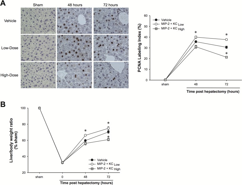
Fig 4. Effects of exogenous MIP-2 and KC treatment on hepatocyte proliferation and liver regeneration after partial hepatectomy.
Wild-type mice were injected intravenously with high doses or low doses of MIP-2 and KC, starting 24 hours after hepatectomy and continued daily. An identical volume of sterile phosphate-buffered saline (PBS) was used as a vehicle control. (A) Hepatocyte proliferation was determined by immunohistochemical staining for proliferating cell nuclear antigen (PCNA) and quantitative analysis of PCNA labeling. Data are mean ± SEM with n = 4–8 per group. *P<0.05 compared to vehicle group. Original magnification was 400X. (B) Liver regeneration was determined by increases in liver mass. Data are mean ± SEM with n = 4–8 per group. *P<0.05 compared to vehicle group.

