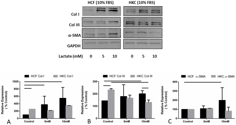
Figure 2. Quantification of western blots of HCF and HKC cell lysates following four week treatment with increasing concentrations of lactate.
(A) Col I expression was significantly up regulated (p < 0.05) in HCFs following 5 and 10 mM exogenous lactate stimulation, (B) Col III was significantly up regulated (p < 0.05) in HCFs following 10 mM exogenous lactate stimulation. Col III expression was significantly higher at HKC-Control compared to HCF-Control (p < 0.05), and (C) α-SMA was significantly up regulated in HCFs (p < 0.05) following 10 mM exogenous lactate stimulation. All gels have been run under the same experimental conditions. n = 3, error bars represent (SEM).

