
Figure 1. CaspaseTracker biosensor system.
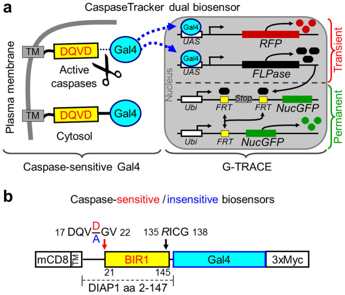
(a) Schematic of the CaspaseTracker biosensor system, which is composed of a plasma membrane anchor (mCD8), a caspase-sensitive natural substrate (derived from DIAP1) fused to the Gal4 transcription factor with a C-terminal 3x-myc tag that translocates to the nucleus upon caspase activation to induce the G-Trace system. (b) Schematic of caspase-sensitive (DQVD) and caspase-insensitive control (DQVA) biosensors with the indicated modifications (NN/GV and D135R)33,34 to prevent biosensor degradation and to prevent caspase inhibition by the biosensor.