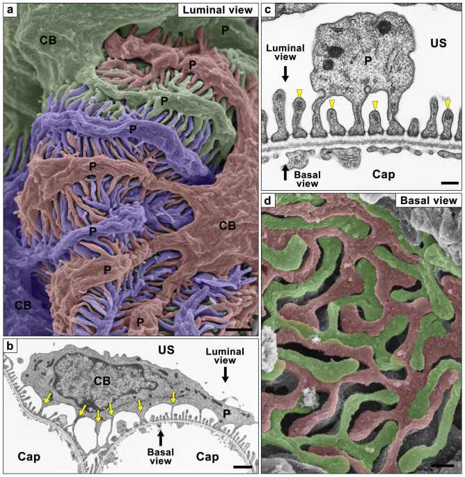
Figure 1. Podocyte subcellular compartments shown by conventional SEM and TEM.
Podocyte are traditionally divided into three kinds of subcellular compartment, cell body (CB), primary process (P) and foot process. (a) Conventional SEM image shows the luminal surface of all three kinds of podocyte subcellular compartments. Three neighboring podocytes are individually colored with blue, green, and red. (b, c) Conventional TEM images also show the three subcellular compartments. The foot processes are predominantly protruding from the primary processes, but some of them are from the undersurface of cell body (yellow arrows in b). The foot processes possess the electron-dense actin bundle within their luminal cytoplasm (yellow arrowheads in c). Directions of luminal and basal views are indicated by black arrows. (d) Alkaline-maceration SEM images clearly show the basal surface of neighboring podocytes. The region in contact with the glomerular basement membrane consists almost entirely of the interdigitating foot processes. Two neighboring podocytes are individually colored with green and red. Cap, capillary lumen. US, urinary space of the Bowman's capsule. Bar scales, 1 μm in (a), (b); 200 nm in (c), (d).

