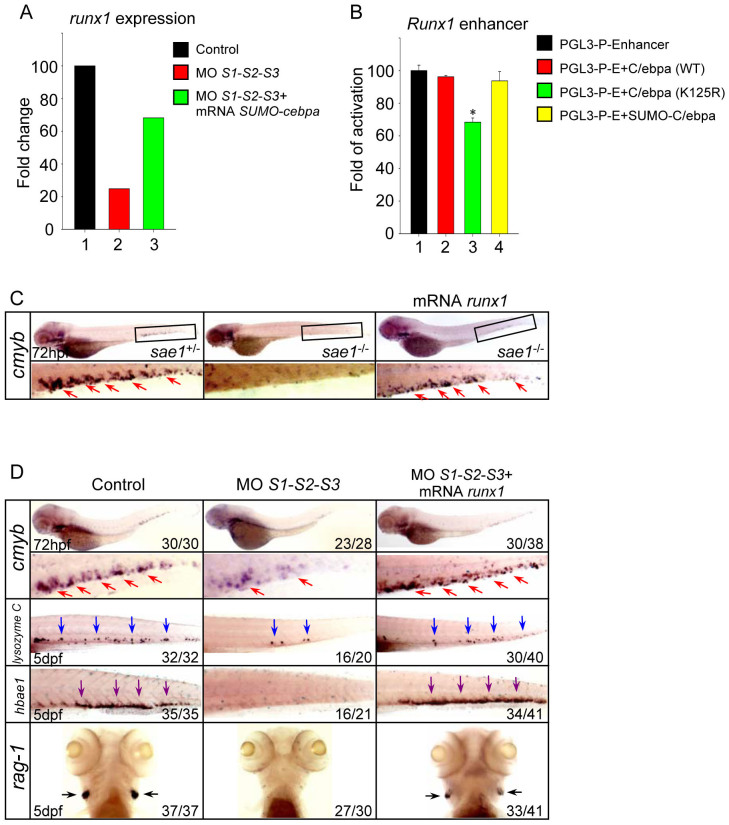
Figure 5. Runx1 is implicated in the hematopoietic defects triggered by SUMO-deficiency.
(A) The relative expression level of runx1 revealed by microarray analysis. The result shown is expressed as fold difference compared with the level (set to 100) detected in control embryos. (B) Luciferase activity assays were performed in 293T cells using the various C/ebpα constructs indicated. The Renilla plasmid was used as an internal control. Data shown are the mean ± SD of three independent experiments. *P < 0.05 by student's t-test. (C) WISH assay of cmyb in the sae1 mutant embryos and siblings at 72 hpf. (D) WISH assay of cmyb at 72 hpf and lysozyme C, hbae1 and rag1 at 5 dpf. Note that runx1 overexpression rescued the hematopoietic defects in the SUMO-deficient embryos. Red, blue, purple and black arrows identify cmyb, lysozyme C, hbae1 and rag1-positive cells, respectively.

