
Figure 2.
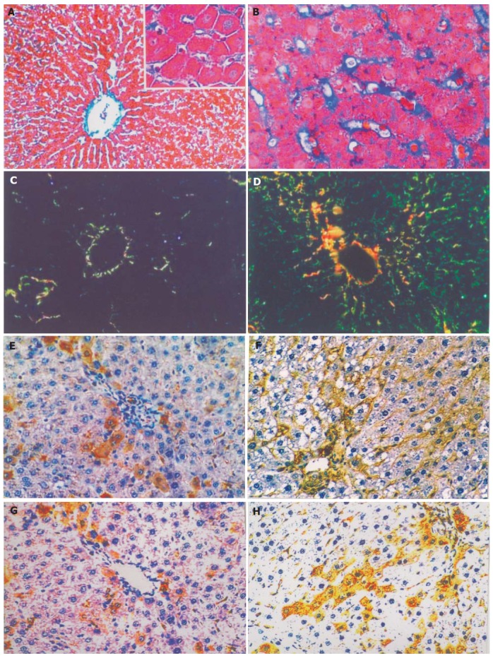
Mallory, Sirius Red and immunohistochemical staining of liver tissue. A: Mallory staining of normal rat liver tissue, ×200 B: Mallory staining of rat alcoholic fibrosis liver tissue, ×400 C: Sirius Red staining of normal rat liver tissue and polarization microscopy, ×200 D: Sirius Red staining of rat alcoholic fibrosis liver tissue and polarization microscopy, ×200 E: Immunohistochemical staining of the first antibody of ColIV of normal rat liver tissue, ×200 F: Immunohistochemical staining of the first antibody of ColIV rat alcoholic liver fibrosis tissue, ×200 G: Immunohistochemistry staining of the first antibody of LN in normal rat liver tissue, ×200 H: Immunohistochemical staining of the first antibody of LN in rat alcoholic liver fibrosis tissue, ×200.