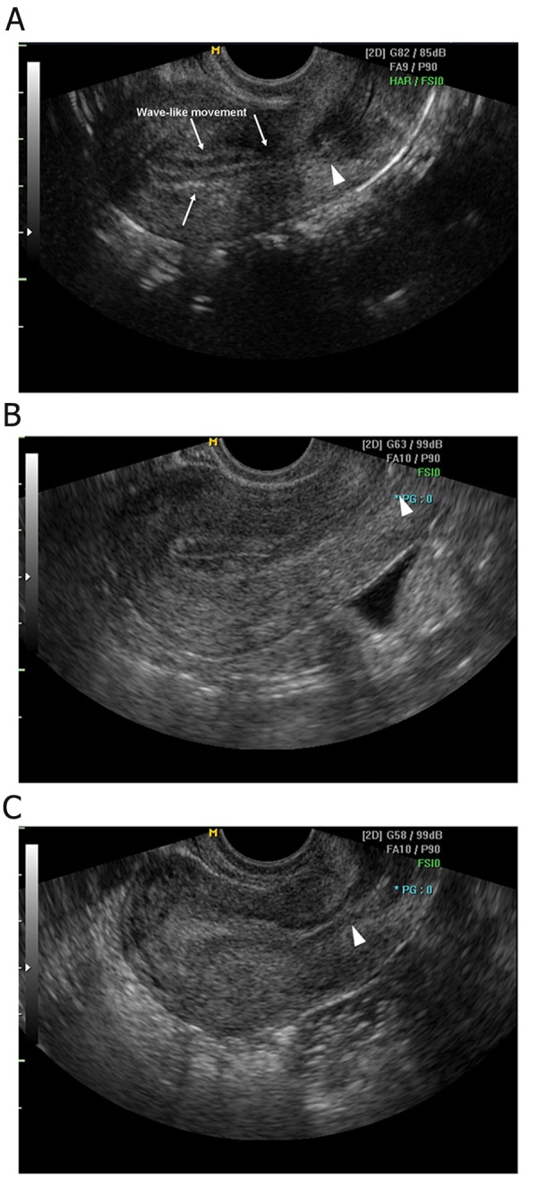
Fig 1.

Ultrasound images of endometrium. Echogenisity was decided by the comparison of myomertial echo. Homogeneously hypoechoic endometrium (A) is defined as a marginal hyperechoic line with a prominent hypoechoic inner layer, in which endometrial wave-like movements are observed. Inhomogeneous endometrium (B) shows an endometrium with a hyperechoic margin and a mottled inner layer, and homogeneously hyperechoic endometrium (C) exhibits an inner layer as hyperechoic as a marginal line. Internal os (arrowheads) can be identified.
