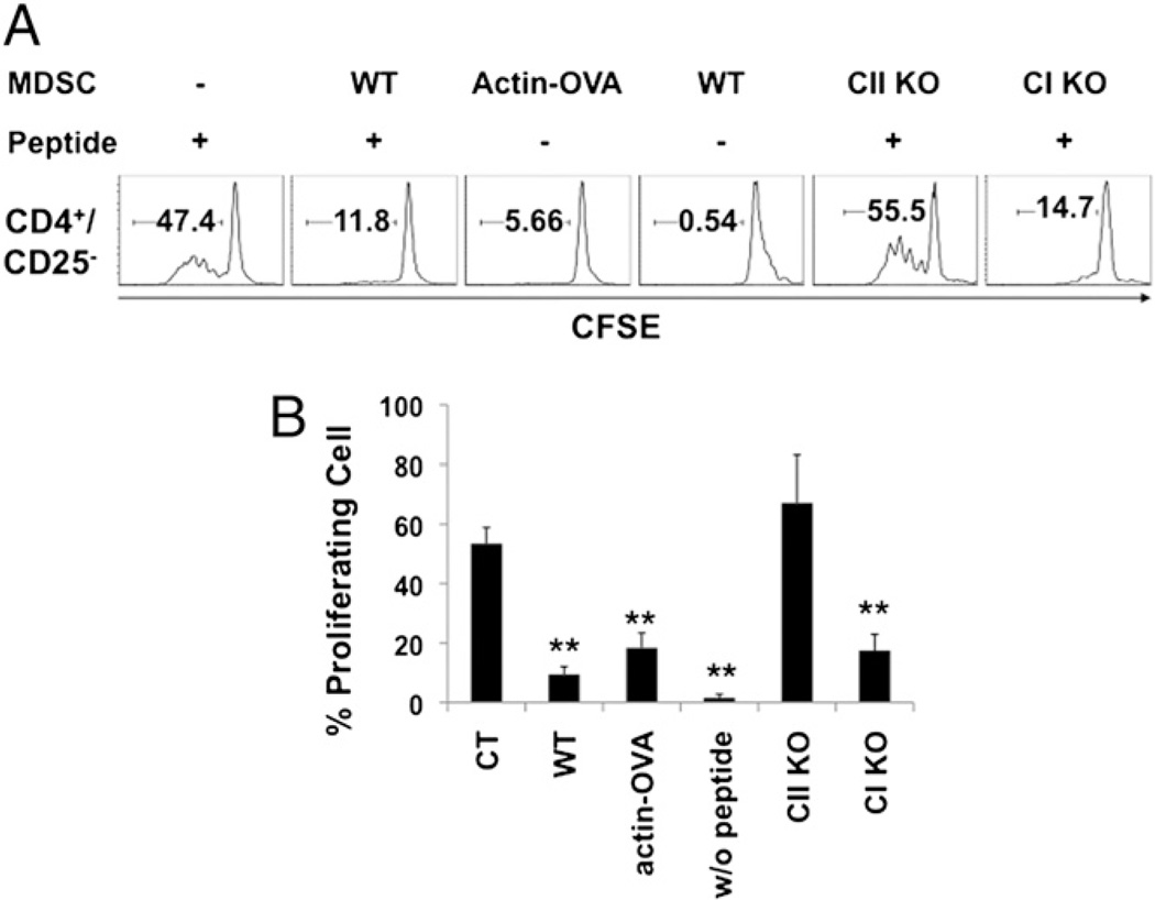
FIGURE 2.
MDSC-mediated suppression of CD4+CD25− T cell proliferation requires MHC class II-restricted Ag presentation. MDSCs were isolated from tumor-bearing WT, actin-OVA transgenic, MHC class I KO, or MHC class II KO mice. Purified CD4+CD25− naive T cells from OT-II transgenic mice were labeled with CFSE and cocultured with MDSCs at a 4:1 ratio or with irradiated naive splenocytes (as APCs) at a 10:1 ratio for 4 d in the presence or absence of exogenously added OVA peptides. Proliferation (CFSE dilution) of CD4+CD25− OT-II cells was assessed by flow cytometry. A, Histograms of CFSE-labeled cells in the population of CD4+CD25− cells from one of the four repeated experiments. B, Statistical analysis of the data from four repeated experiments was performed. Results presented are the average percentage of proliferating CD4+CD25− T cells ± SD from four experiments. **p < 0.001 compared with the control group (i.e., OVA peptide without MDSCs).

