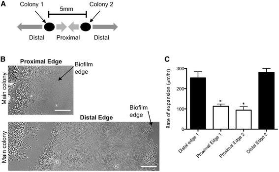
Figure 3.

Self-produced extracellular signals inhibit twitching motility-mediated expansion of P. aeruginosa biofilms. P. aeruginosa PAK inoculated at two adjacent locations 5 mm apart on a solidified media-coated microscope slide results in twitching motility-mediated expansion of two neighbouring biofilms at the interstitial space between the media and coverslip. (A) Assay setup: Two P. aeruginosa PAK colonies inoculated on a nutrient media-coated microscope slide 5 mm apart expand to form two neighbouring interstitial biofilms. (B) Representative phase-contrast microscopy images of P. aeruginosa PAK interstitial biofilms formed at the proximal and distal edges of two neighbouring biofilms after incubation at 37°C for 6 h. Scalebar is 200 μm. Images are representative of the twitching motility zones formed in three independent experiments. (C) The rate of expansion via twitching motility away from the main colony at the proximal and distal edges of two neighbouring interstitial biofilms after incubation at 37°C for 6 h. Mean ± SD is presented from three independent experiments (two-tailed Student’s t-test, *p < 0.05).
