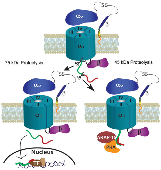
Fig. 1.
Two forms of proteolytic processing of L-type voltage-gated calcium channels. Top: A schematic of an L-type VGCC complex on the plasma membrane. The α1 subunit is the pore-forming subunit and contains four homologous repeats (I–IV). α2δ and β are auxiliary subunits. The red and green regions represent the C-terminus of α1. Two sites of proteolysis are indicated. Bottom left: The 75 kDa cut: the C-terminus of α1 is cleaved from the channel and translocates to the nucleus to interact with transcription factors (TF) and influence gene expression. Bottom right: The 45 kDa cut: the distal C-terminus (red) of α1 is cleaved from the channel and reassocciates with the proximal C-terminus (green) causing inhibition of the channel. AKAP-15 and PKA are docked close to this site. When activated by the β-adrenergic pathway, PKA phosphorylates a site to dislodge the distal C-terminus, relieving the inhibition.

