. 2014 Sep 10;3:e03728. doi: 10.7554/eLife.03728

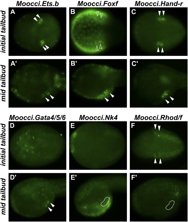
Figure 2. Expression of conserved TVC/heart markers in M. occidentalis embryos.

In situ hybridization (ISH) in M. occidentalis embryos for (A and A′) Moocci.Ets.b, (B and B′) Moocci.Foxf, (C and C′) Moocci.Hand-related (Moocci.Hand-r), (D and D′) Moocci.Gata4/5/6, (E and E′) Moocci.Nk4, (F and F′) Moocci.Rhod/f. ISH was performed on initial tailbud (AF) and mid tailbud (A′F′) stage embryos. Solid arrowheads indicate definitive expression in TVCs. Dotted arrowheads indicate potential expression of Moocci.Foxf in initial tailbud, obscured by strong epidermal expression. Dotted outline indicates probable position of TVCs, not visible due to lack of mRNA hybridization signal. Initial tailbud embryos were imaged ventrally or dorsally, while tailbud embryos were imaged laterally.

DOI: http://dx.doi.org/10.7554/eLife.03728.008

Figure 2.

Figure 2—figure supplement 1. Ets.b expression in B7.5 of M. occidentalis.
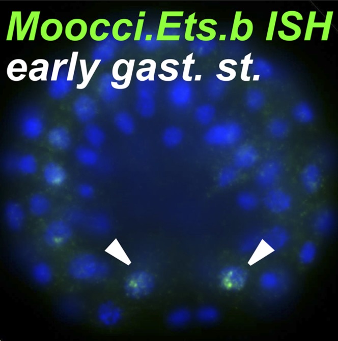
Figure 2—figure supplement 1.

In situ hybridization (ISH) for Moocci.Ets.b in 110-cell st/early gastrula embryo, showing expression (green) in the B7.5 blastomeres. Nuclei are counterstained with DAPI (blue). Embryo was imaged from a vegetal view, with anterior to the top.
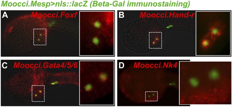
Figure 2—figure supplement 2. In situ hybridizations reveal TVC gene expression in M. occidentalis.

Figure 2—figure supplement 2.

Mid-tailbud M. occidentalis embryos electroporated with Moocci.Mesp>nls::lacZ, assayed with β-galactosidase immunodetection (IHC, green) couple to in situ hybridization (ISH, red) for (A) Moocci.Foxf, (B) Moocci.Hand-r, (C) Moocci.Gata4/5/6, and (D) Moocci.Nk4. Expression of these genes was detected in the TVCs, with the possible exception of Nk4. All embryos are oriented with anterior to the left.
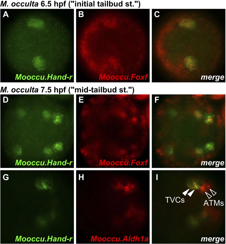
Figure 2—figure supplement 3. Hand-r and FoxF co-expression reveals TVCs of M. occulta.

Figure 2—figure supplement 3.

Coronal view of M. occulta embryos at (AC) 6.5 hpf (equivalent to the initial tailbud stage of tailed M. oculata) and (DI) 7.5 hpf (equivalent to the mid-tailbud stage). Embryos were assayed for expression of Mooccu.Hand-r (green—A, D, G) and Mooccu.Foxf (red—B, E) or Mooccu.Aldh1a (red—H) by double in situ hybridization. Merged green and red channels show co-expression in TVCs (C, F, I). Mooccu.Foxf expression in epidermis seen in F suggests position of TVCs in ventro-lateral regions of the trunk similar to what is observed in M. occidentalis and C. intestinalis. Mooccu.Aldh1a-expressing cells immediately posterior to TVCs are likely the anterior tail muscles. Anterior is to the left in all panels.
Figure 2—figure supplement 4. Aldh1a expression in M. occidentalis embryo.
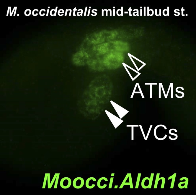
Figure 2—figure supplement 4.

In situ hybridization for Moocci.Aldh1a showing expression in TVCs and ATMs in a mid-tailbud embryo.