. 2014 Sep 10;3:e03728. doi: 10.7554/eLife.03728

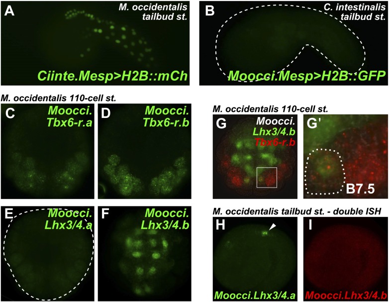
Figure 4. Developmental system drift of Mesp regulation between C. intestinalis and M. occidentalis.

(A) M. occidentalis tailbud embryo electroporated with a Ciinte.Mesp>H2B::mCherry reporter construct. Weak reporter gene expression was observed in the B7.5 lineage and occasionally in other territories including B-line mesenchyme and tail muscle cells, and A-line neural plate derivatives. (B) C. intestinalis tailbud embryo electroporated with Moocci.Mesp>H2B::GFP reporter. No fluorescence was seen in any cells, indicating complete lack of activity of Moocci.Mesp enhancer in wild-type C. intestinalis embryos. (C) In situ hybridization (ISH) for Moocci.Tbx6-r.a, (D) Moocci.Tbx6-r.b, (E) Moocci.Lhx3/4.a, and (F) Moocci.Lhx3/4.b in 110-cell stage embryos. (G) Double ISH in 110-cell stage embryo reveals co-expression of Moocci.Lhx3/4b (green) and Moocci.Tbx6-r.b (red) exactly in the B7.5 cells of M. occidentalis. (G′) Magnified view of inset in (G). (H) Double ISH for Moocci.Lhx3/4.a (green) and (I) Moocci.Lhx3/4.b (red) in a mid-tailbud embryo. Moocci.Lhx3/4.a but not Moocci.Lhx3/4.b is expressed in motor ganglion neurons (arrowhead).

DOI: http://dx.doi.org/10.7554/eLife.03728.017

Figure 4.

Figure 4—figure supplement 1. Weak and leaky expression of Ciinte.Mesp reporter in M. occidentalis embryos.
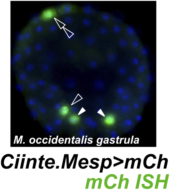
Figure 4—figure supplement 1.

In situ hybridization for mCherry mRNA (green) in a M. occidentalis early gastrula stage embryo electroporated with Ciinte.Mesp>mCherry. Nuclei counterstained with DAPI (blue). Embryo is viewed vegetally, anterior to the top. Expression in B7.5 blastomeres (solid arrowheads) was observed in 42% of embryos. Ectopic expression in other B-line cells (hollow arrowhead) was seen in 24% of embryos. Ectopic expression in A-line neural precursors (hollow double arrowhead) was seen in 8% of embryos. In contrast, in situ hybridization revealed that Moocci.Mesp reporter construct is expressed in the TVCs in 60% of electroporated M. occidentalis embryos, with 0% embryos showing any ectopic reporter gene expression (data not shown, see Figure 1D for example). n = 50 embryos for each construct.
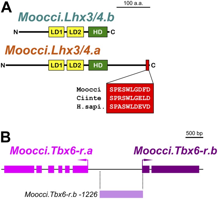
Figure 4—figure supplement 2. Configuration of Lhx3/4 protein domains and Tbx6-r locus in M. occidentalis.

Figure 4—figure supplement 2.

(A) Schematic of Moocci.Lhx3/4.b and Moocci.Lhx3/4.a proteins. LIM domains (LD1 and LD2) are in yellow, and the homeodomain (HD) is in green. Moocci.Lhx3/4.a retains a more extensive C-terminus including a highly conserved motif (highlighted in red) of unknown function. Alignment to Lhx3/4 orthologs from C. intestinalis (Ciinte) and humans (H.sapi.) is shown in inset. (B) Schematic representing the Tbx6-related locus in M. occidentalis, showing head-to-head configuration of Tbx6-r.a and Tbx6-r.b. Exons are represented by thick blocks. Tbx6-r.a is encoded by 6 exons, while Tbx6-r.b is encoded by only 2 exons. The region corresponding to the Moocci.Tbx6-r.b driver used in this study is shown in periwinkle.
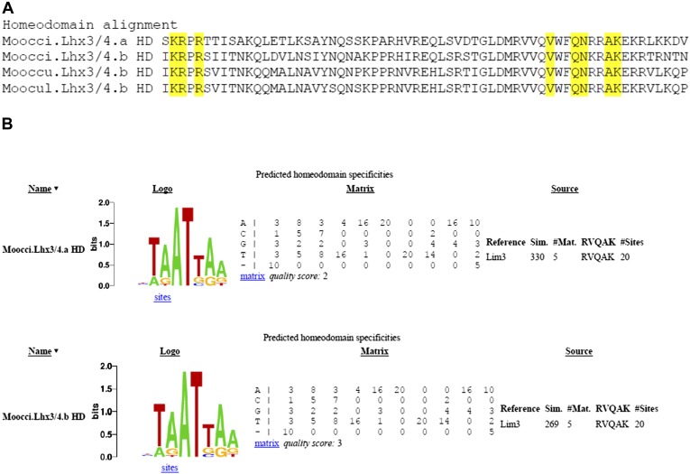
Figure 4—figure supplement 3. Divergent Molgula Lhx3/4.b homeodomains are not predicted to have altered DNA binding specificities.

Figure 4—figure supplement 3.

(A) Alignment of homeodomains (HDs) from a set of Molgula Lhx3/4 family proteins, with HD recognition positions highlighted in yellow. HD recognition positions are invariant while intervening sequence is highly diverged between Lhx3/4.a and Lhx3/4.b. (B) Logos and matrices for predicted homeodmain specificities for Moocci.Lhx3/4.a (top) and Moocci.Lhx3/4.b (bottom), generated by the Homeodomain Specificity Prediction web page (http://stormo.wustl.edu/cgi-bin/flyhd/hd_pred.cgi; Noyes et al., 2008). The two predicted binding specificities are identical to one another, due to perfect conservation of the HD recognition positions.