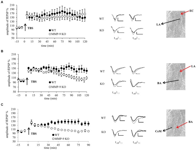
Figure 1.
Genetic inhibition of MMP-9 results in destabilization of LTP in the central and basal but not in the lateral amygdala. (A) fEPSP in the EC–LA amygdala pathway was similar in slices from mice lacking functional MMP-9 gene (MMP-9 KO, open circles n = 6) and control animals (WT, filled circles, n = 5). (B) fEPSP evoked in the LA-BA pathway in slices from MMP-9 KO mice (open circles, n = 7) within first 70 min had the same magnitude as LTP in slices from control animals (WT, filled circles, n = 7); however afterwards it went down to the baseline level. (C) fEPSP induced in the BA-CeAm pathway in slices from MMP-9 KO mice (open circles, n = 7) had the same amplitude as LTP evoked in control slices (filled circles, n = 7) within first 30 min after induction. Then, LTP in MMP-9 KO slices gradually decreased to the baseline level. Left panels show graphs with time course of maximal EPSP amplitudes normalized to baseline. Black arrows mark the time of application of TBS stimulation. Error bars represent SEM. Middle panels show exemplary traces of fEPSP recorded 10 min before (black) and 15 and 90 min after (gray) induction of LTP. Scale bars = 0.2 mV and 5 ms. Right panels present photographs of mouse amygdala (Nissl staining) with positions of stimulating (red arrow) and recording (black arrow) electrodes.

