Figure 1.
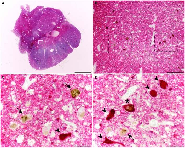
Coronal section through the ventral mesencephalon showing the obtained dual stain for α-synuclein (TH; brown) and tau (red). At the level of the substantia nigra pars compacta (SNc), up to three different types of melanin-containing neurons were observed, comprising (i) brown-stained neurons containing Lewy bodies (LBs; arrows), (ii) red-stained neurons with tau immunoreactivity (arrowheads); and (iii) neurons showing both LBs and tau deposits (asterisks). Scale bar is 4,000 μm in (A); 200 μm in (B) and 50 μm in the high-magnification insets shown in panels (C) and (D).

