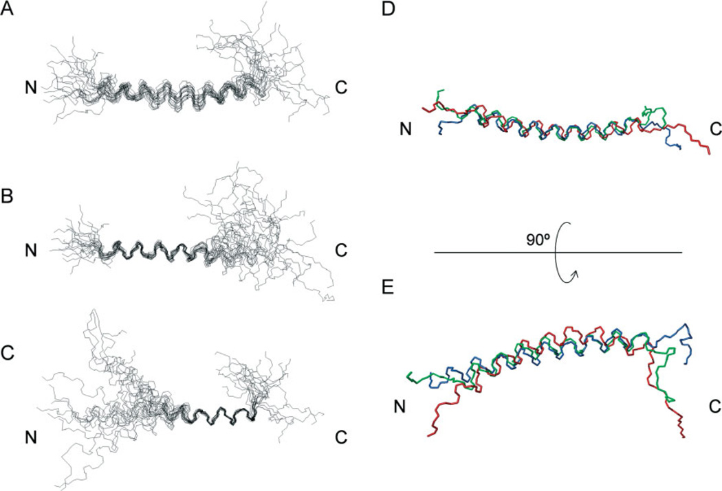
Figure 4.
The tertiary structure of M. sexta moricin in 100% CD3OH. (A)–(C) superimposition of the backbones of 20 lowest energy structures of M. sexta moricin best fitted to residues 5–36 (A), to residues 5–22 (B), and to residues 23–36 (C). (D) Superimposition of the average, lowest energy structures of B. mori (blue), S. litura (green) and M. sexta (red) moricins; (E) the same as (D) but rotated 90° around the horizontal axis.

