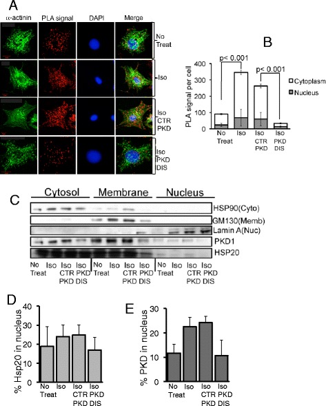
Figure 3.

Hsp20 may act as a nuclear chaperone for PKD1. A and B. A novel proximity ligation assay was used to visualize and quantify PKD1 nuclear entry in neonatal cardiac myocytes. Images are shown as maximum projections of z-stacks of confocal images. PLA signals indicating Hsp20-PKD1 complex formation were quantified using the analyse particles plugin of ImageJ software. For all experiments, quantifications were performed from at least 12 images and expressed as mean number of signals per cell. C, D and E. Cell fractionation techniques were used to visualize and quantify the cellular distribution profile of PKD1 and Hsp20 from cardiac myocytes. The disruptor peptide (PKD DIS) prevented ISO-induced nuclear translocation of Hsp20-PKD1 complexes, compared to control peptide (CTR PKD). Data representative of n = 3 independent experiments.
