. 2014 Oct 8;3:e04187. doi: 10.7554/eLife.04187

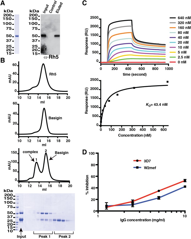
Figure 1. Production of functional recombinant PfRh5.

(A) Purified recombinant PfRh5 was analysed by SDS-PAGE analyses and by erythrocyte binding assays. (B) Formation of the PfRh5–basigin complex was monitored by size-exclusion chromatography. The chromatographic profiles are shown for PfRh5 (panel 1), basigin (panel 2), and the PfRh5-basigin complex (panel 3). The fractions eluted from the column in panel 3 were analysed by SDS-PAGE. (C) The binding affinity of the recombinant PfRh5 to human basigin was measured by SPR on Biacore 3000 with the basigin coupled to a sensor chip. (D) In vitro growth inhibition assays were performed to assess the abilities of the polyclonal antibodies to the recombinant PfRh5 in blocking P. falciparum parasite invasion into erythrocytes.

DOI: http://dx.doi.org/10.7554/eLife.04187.003

Figure 1.

Figure 1—figure supplement 1. Production of full-length PfRh5.

Figure 1—figure supplement 1.

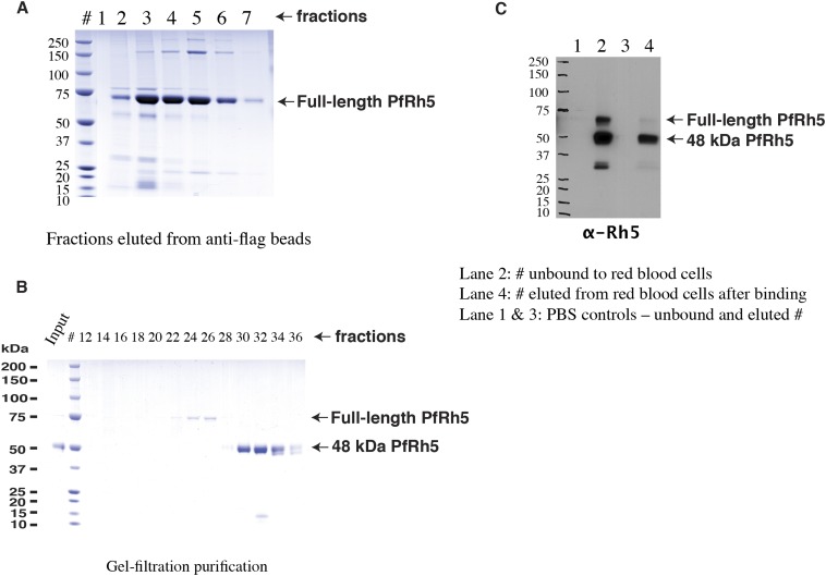
(A) SDS-PAGE analyses of the FLAG-tagged recombinant full-length PfRh5 eluted from an anti-FLAG affinity column. (B) Size-exclusion chromatography analyses of the anti-FLAG bead affinity purified PfRh5. The fractions eluted from the affinity column were pooled, concentrated, and stored at 4°C for 2 days before the analyses. (C) Red blood cell binding assay with PfRh5 purified by anti-FLAG bead affinity chromatography. Both full-length protein and the breakdown fragments bound red blood cells with a 48-kDa species having the highest affinity.
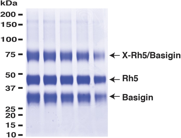
Figure 1—figure supplement 2. PfRh5 and human basigin form a 1:1 complex.

Figure 1—figure supplement 2.

PfRh5-C and basigin were cross-linked with EDC in the presence of NHS. The mixture was then analysed on a SDS-PAGE gel and the band of the crosslinked complex was excised for trypsin/chymotrypsin digestion followed by mass spectrometric analysis.