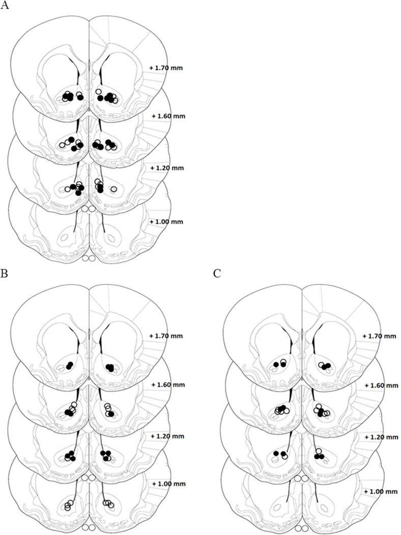
Figure 6. Location of the microinjection guide cannula in the nucleus accumbens from experiments 5 and 6.
A. Illustration showing the location of AMPA microinjection cannula tips in the Core. The open circles represent cannula placements in non-stress group and open filled circles depict placements in stress group. B-C Illustration showing the location of DMSO and CNQX microinjection cannula tips in the Core. The open circles represent cannula placements in non-stress group and open filled circles depict placements in stress group.

