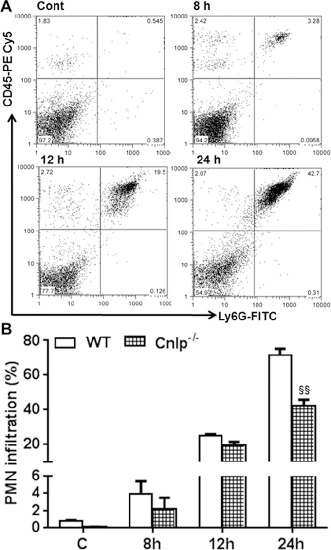
Figure 8.

Cnlp−/− mice exhibit reduced PMN infiltration at the early stages of infection. Eyes (n = 8) of WT (C57BL/6) and Cnlp−/− mice (B6 background) were infected with S. aureus (5000 CFU/eye of strain RN6390). Polymorphonuclear neutrophil infiltration was determined using flow cytometry (A), as described in the legend to Figure 4. Bar graph shows cumulative quantitative data from three independent experiments (B). Student's t-test was used for statistical analysis, and comparisons were made between WT versus Cnlp−/− (§); (§P < 0.05; §§P < 0.005).
