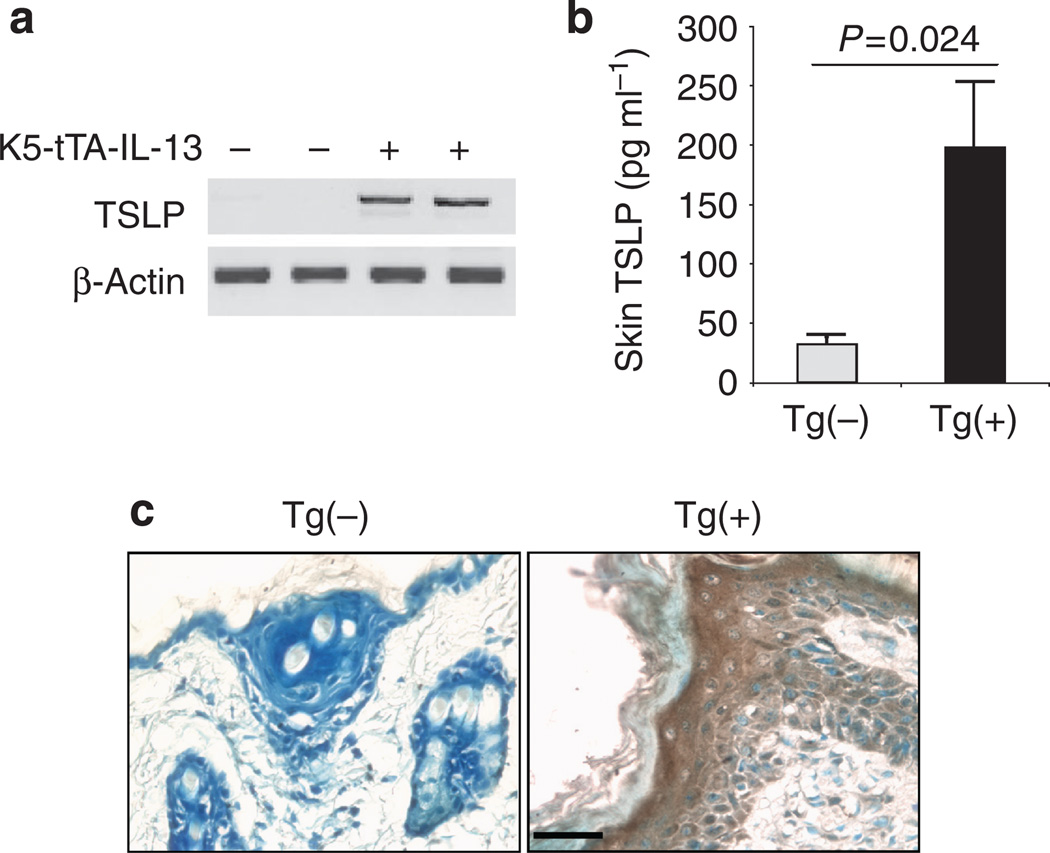
Figure 5. Upregulation of TSLP expression in the skin.
(a) RT–PCR for TSLP mRNA in the skin. (b) TSLP protein in the skin extracts (n = 6 for each group). (c) IHC identification of cell types expressing TSLP. Skin sections from Tg(+) and Tg(−) mice were stained using anti-TSLP. Bar = 50 µm. Shown are representative slides of three pairs of stained samples.

