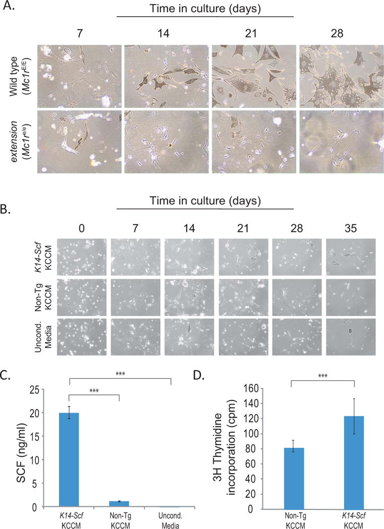
Figure 2.

Progressive purification of primary melanocytes is facilitated by stem cell factor. A) Phase contrast photographs of primary melanocytes from Mc1r-intact (wild type) or –defective (extension) strains (magnification 100×). Note that cultures become pure after 3–4 weeks in culture, as determined by morphology (dendricity). Also note that melanocytes from Mc1r-intact (wild type) strains are much larger and more robustly pigmented than their Mc1r-defective counterparts. B) Comparison of cultures of Mc1r-defective (extension) melanocytes grown in complete primary melanocyte growth media made with media conditioned from either K14-Scf transgenic primary keratinocytes (upper panels) or non-transgenic primary keratinocytes (middle panels) or grown in unconditioned media (lower panels). Note that cells grew better with conditioned media from K14-Scf transgenic keratinocytes. C) Quantification of the amount of soluble SCF in Complete Primary Melanocyte Growth Media made with conditioned supernatants from K14-Scf transgenic primary keratinocytes, non-transgenic primary keratinocytes or unconditioned media as indicated. SCF quantification was performed by ELISA (RayBiotech Systems) and statistical significance (p <0.01) is indicated (***). D) 3H-thymidine incorporation of purified Mc1re/e (extension) melanocytes grown in complete primary melanocyte growth media made either with keratinocyte conditioned media from non-transgenic primary keratinocytes or K14-Scf transgenic keratinocytes as labeled. Melanocytes grown in media containing the K14-Scf conditioned media incorporated a significantly greater amount of 3H-thymidine than melanocytes grown in non-transgenic conditioned media (p < 0.018).
