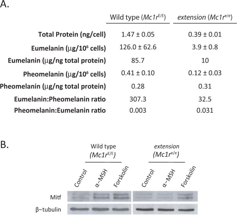
Figure 3.

Determination of melanin levels and MSH responsiveness of primary melanocytes. A) Total protein content of purified primary melanocytes grown for 4–5 weeks in culture was determined by the BCA Protein Assay (Thermo Fisher Scientific). Similarly, eumelanin and pheomelanin were quantitatively analyzed by HPLC based on the formation of pyrrole-2,3,5-tricarboxylic acid (PTCA) by permanganate oxidation of eumelanin and 4-amino-3-hydroxyphenylalanine (4-AHP) by hydriodic acid reductive hydrolysis of pheomelanin, respectively as described (Ito and Wakamatsu 2003). B) Western analysis of Mitf expression in response to 100 nM α-MSH or 100 μM forskolin with β-tubulin loading control.
